Joint arthroscopy is a minimally invasive surgical procedure used to diagnose and treat problems inside a joint. Instead of a large incision, the surgeon makes small keyhole incisions and inserts a thin instrument called an arthroscope that has a camera and light. The image is displayed on a monitor, allowing the surgeon to see the inside of the joint clearly.
Common Joints Treated
Arthroscopy is commonly performed in:
Knee joint
Shoulder joint
Ankle joint
Hip joint
Wrist
Elbow
Conditions Treated with Arthroscopy
Meniscus tear
Ligament injuries (ACL/PCL)
Cartilage damage
Loose bodies in the joint
Synovitis
Rotator cuff tear (shoulder)
Basic Procedure Steps
Small incisions (portals) are made around the joint.
Arthroscope is inserted to visualize the joint.
Sterile fluid is pumped to expand the joint for better viewing.
Special instruments are inserted through other portals to repair or remove damaged tissue.
Incisions are closed with sutures or sterile strips.
Advantages of Arthroscopy
Smaller incisions
Less pain
Minimal tissue damage
Shorter hospital stay
Faster recovery
Better cosmetic results
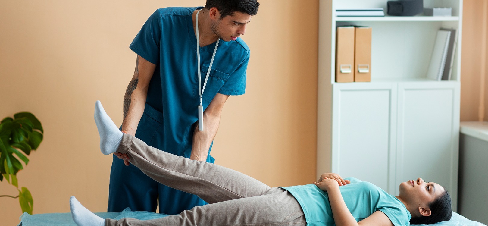
Recovery
Many procedures are day-care surgeries.
Physiotherapy is often required.
Return to activity may range from a few weeks to several months, depending on the procedure.
Arthroscopy has become a standard technique in modern orthopaedics, especially for knee and shoulder joint conditions because it allows precise treatment with minimal surgical trauma.